
A chromatogram can show that one species dominates a sample. It cannot say that the species is the intended molecule. Identity is a separate question, answered with other instruments and, ideally, with several of them. This article walks through the usual steps. Terms are defined in the The testing glossary: 14 lab terms in plain English.
Purity and identity are different questions
A sample can be very clean and still be the wrong molecule. Chromatography ranks how much of the detected material sits in one peak. Identity needs evidence about mass, sequence and structure.
Why one method is not enough
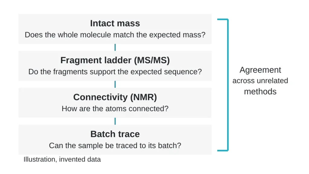
Laboratories use orthogonal methods, which means methods built on different physical principles. Liquid chromatography separates by how strongly molecules interact with a column. Mass spectrometry measures mass-to-charge ratio. Tandem mass spectrometry breaks selected ions apart and measures the pieces. NMR probes the chemical surroundings of atoms.
One method can mislead or be misread. Several unrelated methods that all agree are much harder to explain away. Peptide reference-standard programs reflect this: identity testing in a review from the United States Pharmacopeial Convention combines NMR, mass spectrometry and chromatography, and the authors note that features such as chiral or isobaric amino acids may need additional techniques (McCarthy et al., 2023).
Step 1: intact mass
The first check compares the measured mass of the whole molecule with the theoretical mass calculated from its formula. High-resolution instruments such as quadrupole time-of-flight and Orbitrap systems report the difference as a mass error in parts per million:
error in ppm = (observed minus theoretical) divided by theoretical, times one million
Invented example: if the observed mass is higher than the theoretical mass by four millionths of its own value, the error is plus 4.0 ppm. Agreement at that level supports the proposed formula.
It does not settle the arrangement of atoms. Different structures can share the same overall mass, which is why the next steps exist.
Step 2: sequence by tandem mass spectrometry
In tandem mass spectrometry the instrument selects one ion, fragments it under controlled conditions and records the fragments. Neighboring fragment peaks in a series differ by the mass of one amino acid residue, so the series can be read like a ladder.
Invented example: if neighboring peaks in a series sit 57.02 apart and the next pair sits 71.04 apart, the first gap matches a glycine residue and the second matches an alanine residue. Reading the full ladder gives the order of residues.
Laboratories summarize the result as sequence coverage, the share of the expected sequence supported by fragment evidence. One published case study of an unknown synthetic protein reported a de novo sequence coverage of 70 percent, similar to what searching a sequence database achieved (Pino et al., 2019). A coverage figure therefore needs context: which regions are covered, and whether modified regions are among them.
Step 3: linkages and structure
Some molecules contain a linkage between two residues, such as a disulfide bond between two cysteines. Two samples can share a sequence and still differ in how they are linked, and mass alone may not separate them.
Linkages also complicate fragment reading. In a published study of a disulfide-containing synthetic peptide, the linkage made degradation products hard to characterize, so the authors chemically reduced it before liquid chromatography with high-resolution tandem mass spectrometry, which allowed eleven degradation products to be assigned (Chawathe and Sharma, 2025).
NMR addresses connectivity directly. A one-dimensional proton spectrum lists hydrogen environments but often overlaps heavily for a molecule of this size. A COSY experiment adds a second axis and shows which protons are coupled through nearby bonds. An HSQC experiment links each proton signal to the carbon it is attached to. Together they let an analyst assign signals and test whether the connectivity fits the proposed structure. NMR needs more material than mass spectrometry and relies on expert assignment.
Step 4: purity, content and aggregates alongside identity
Identity evidence sits next to other measurements. Reversed-phase chromatography gives purity. A content assay gives milligrams of target in the vial, as described in CHNS, liquid chromatography or LC-MS: which test answers which question?. Size-exclusion chromatography separates by apparent size and can reveal dimers and larger aggregates that a reversed-phase method may not distinguish. Endotoxin is a separate assay, covered in EU per vial: how to read an endotoxin number.
Each of these answers a different question. A pure sample can be underfilled, a correctly filled sample can contain more impurity than expected, and a clean chromatogram says nothing about bacterial endotoxin.
Step 5: traceability
Independent reports only add up if they describe the same material. Look for the same lot identifier, sample identifier and date range across the mass spectrometry, NMR and chromatography documents. Without that link, three good reports could describe three different lots.
A reader’s checklist
- Is the intact mass given with a mass error, and is that error small?
- Was the sequence checked by fragmentation, and which regions have coverage?
- If the molecule has a linkage, which method addresses it?
- Do the methods rest on different principles?
- Does every report carry the same lot identifier?
More questions to put to a laboratory are in The questions a good testing lab answers without flinching.
Sources
- McCarthy D, Han Y, Carrick K, Schmidt D, Workman W, Matejtschuk P, Duru C, Atouf F. Pharmaceutical Research, 2023, volume 40(6), pages 1317-1328. PubMed ID 36949371. DOI 10.1007/s11095-023-03493-1. Descriptor: peptide reference standards, identity testing by NMR, mass spectrometry and chromatography, and the note on chiral and isobaric residues.
- Pino L, Lin A, Bittremieux W. Journal of Proteome Research, 2019, volume 18(11), pages 3936-3943. PubMed ID 31556620. DOI 10.1021/acs.jproteome.9b00384. Descriptor: case study of de novo sequencing of an unknown synthetic protein, 70 percent sequence coverage.
- Chawathe A, Sharma N. Analytical and Bioanalytical Chemistry, 2025, volume 417(19), pages 4311-4329. PubMed ID 40518483. DOI 10.1007/s00216-025-05944-7. Descriptor: study of a disulfide-containing synthetic peptide, chemical reduction before UHPLC high-resolution tandem mass spectrometry.
Want the catalog? Register free